A Skull Fracture Lawyer
This site is intended to benefit:
- Victims of skull fractures
- Attorneys helping skull fracture victims
This site teaches:
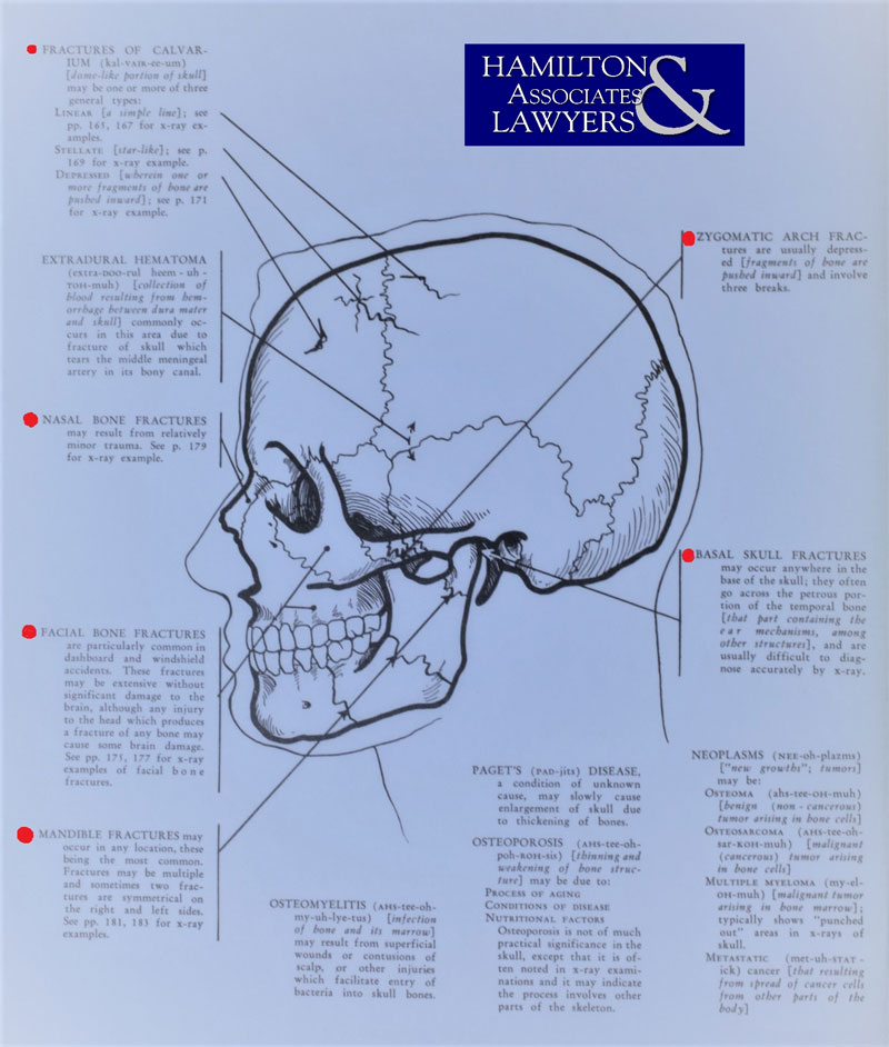
- The Types of Skull Fractures
- How Common are Skull Fractures
- What To Look For in various types of skull fractures
- How are Skull Fractures Treated
- The Most Common Causes of each type of broken skull bone
- How to Diagnose and Identify the Types of Skull Fractures
- Complications from a skull fracture
The Skull Fracture Lawyers at Hamilton and Associates have targeted experience and specific success in helping victims of skull fractures. The content here has been created to help others who have been affected by the traumatic accidents that result in skull fractures.
Open versus Closed Skull Fractures
There are two basic kinds of skull fractures; known as open and closed. The distinction is simple. If the injury involves a piece of the bone in your head sticking through the skin, you have an “open” fracture. Otherwise, you have a “closed” bone break within your skull.
Diastatic Bone Breaks
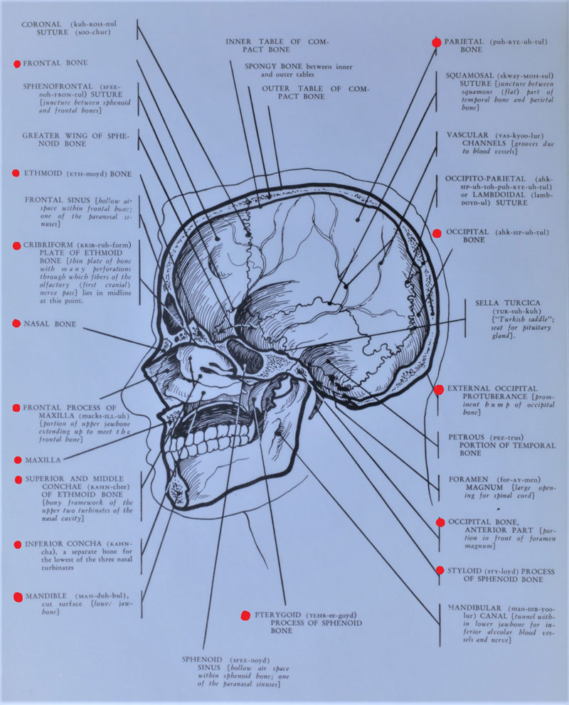
Each person’s head is made up of various bones that are fused together. The lines along your skull that fuse these together are called “suture lines.” Blunt trauma to the head can sometimes fracture (break) these diastatic sutures lines. Diastatic breaks are sometimes called “growing fractures” as children, especially infants, have softer skulls along these dividing lines and are more susceptible to diastatic injury.
Diastatic skull fractures are typically caused by a force coming into the impact the head over a large area of space. Diastatic fractures are often seen resulting from an impact with the side of a vehicle or hitting a wall.
Treatment usually requires a full hospital admission for observation and monitoring. One may or may not expect surgery, depending upon the severity and location of the growing fracture.
Depressed Skull Fracture
A “Comminuted” bone break is one where the skull fractures into multiple shards or pieces. A “Depressed” skull fracture can be seen because the head will appear indented. These can be especially serious if the indention pushes into the brain cavity. Depressed skull fractures are nearly always comminuted.
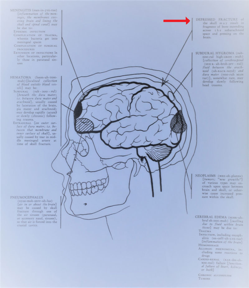
Depressed skull fractures are often called “ping pong” breaks. They are opposite of a diastatic fracture in that they result from a severe and direct impact over a very small area of the head. The skull bones are then broken into multiple pieces and pushed toward the brain. Depressed skull fractures typically happen in the front and top part of the head. These areas, such as the front lobe and parietal lobe, are where the skull bone is thinnest and most susceptible to direct impact.
Basic depressed skull breaks are treated expectantly. This is even so for an infant victim (who is otherwise intact). These bone breaks tend to heal well and work themselves out over time. They often do not even need a formal treatment through elevating the indention.
DANGER: Cerebral spinal fluid can begin to leak into the skull and build pressure. Surgery is needed in these instances. Other types of depressed skull fracture surgeries use the fragments of donors or synthetic fragments to protect the brain. Expect a prescription for rehabilitation after a depressed skull fracture occurs.
Basilar Skull Fractures
These are broken bones in the head at the base of one’s skull. One typically sees basilar fractures in the following areas:
- Eyes
- Ears
- The back near the spine
- Nose
There are three main types of basilar skull fractures. They are:
- Comminuted “shattered into multiple pieces”
- Green Stick breaks “not having broken through the entire bone”
- Linear fractures “straight line through the bone”
Conservative management treatment is typically the initial prescription of the medical treatment provider. Often antibiotics are not prescribed. Expect the doctors to be looking for cerebral spinal fluid dripping from the ears or nostrils. This may require surgery.
Mid Facial Skull Fractures
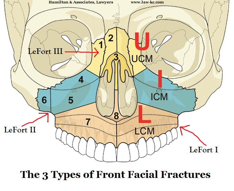
The French surgeon, Dr. Rene Le Fort created a classification system for mid face skull fractures in 1901. This was done in his seminal publication: Étude expérimentale sur les fractures de la mâchoire supérieure. Seventeen years later, Dr. Le Fort followed up his publication with the second article, Les Projectiles inclus dans le mediastin. This is translated as “The Projectiles Included in the Mediastinum.”
All mid facial fractures go through the pterygoid plates. They are known as Le Fort I, Le Fort II and Le Fort III fractures. We examine them as follows:
Mid Facial Breaks – Le Fort I – Horizontal fractures
A horizontal break of the skull midway down the face is known as a Le Fort I mid facial fracture. This is the break of the maxillary, which is the area above your teeth and above your palate. Le Fort I fractures can extend into your head and above the top of your mouth. In essence, these bone breaks separate your dental arch from the part of your skull around your eye socket and occur between your cheekbones and sinuses.
They go by other names other than a Le Fort I fracture, such as:
- Floating palate fracture
- Guerin fracture
- Horizontal maxillary fracture
- Horizontal LeFort fracture
- LeFort I fracture
There have been difficulties in diagnosing LeFort fractures Symptoms of these horizontal skull breaks are as follows:
- Blurred vision
- Bleeding
- Tenderness
- Numbness
- Malocclusion
- Nasal obstruction
- Pain at injury site with a headache
- Swelling over the lips
Mid Facial Fractures (LeFort II – Pyramid Break)
LeFort II fractures are often called pyramidal maxillary fractures. This is because the bone around this area is roughly shaped liked a triangle at the front of one’s face.
Causes of LeFort II pyramidal fractures are typically a direct blow to the lower part or middle maxilla. These triangular bone breaks cause the part of the skull on the front of your face to be detached at the upper jawbone. These breaks may extend across your cheeks and eye sockets to either side of your face.
Between 10 and 20 percent of all broken face bones are LeFort II fractures. These often render the victim unconscious. The dangerous part of a LeFort II fracture is suffocation. Expect treatment to immediately include assistance in breathing, with intubation being necessary in some cases. Immediate treatment providers must also look for foreign objects in the throat, mouth, and nose as the broken bones or other objects from the impact can obstruct the victim’s airway. Another complication to breathing is that LeFort II pyramidal fractures often cause large amounts of bleeding which can fill up or obstruct the lungs.
With LeFort II pyramidal fractures, a victim may also suffer brain swelling and swollen jaws are nearly ubiquitous. A LeFort II fracture is also commonly treated along with an injury to the eye socket and the nose.
Mid Facial Fracture – LeFort III – Broken Jawbone
LeFort III facial fractures are often known as transverse fractures or cranial facial dysfunction breaks. The causes of LeFort III fractures are typically a direct impact with the bridge of your nose or upper maxilla. This causes the entire portion of the facial bones to become separated from the rest of the skull.
LeFort III fractures (though rare) are more serious. Surgery is frequently a necessity. Also, these are followed up with orthopedic restructuring of the face itself. LeFort III surgeries often include a frontal bar, titanium plates, and screws being surgical implanted in the face.
Linear Skull Fractures
The most common kind of fractures to a person’s skull is a linear break. These are associated with 70 percent of all broken head bones. Surgery is often not needed if it is not displaced, depressed, distorted, or splintered. A linear skull fracture is relatively simple compared to the other head injuries.
Mandibular Skull Fracture – Broken Lower Jawbone
Extreme pain is what you should expect with a mandibular skull fracture. These broken jawbones are also known to cause dysfunction of the movement and appearance of the mouth. It is important to have any deformity and dysfunction in these areas treated by a medical professional early in the process. The geometry of the mandible and temporomandibular joints (TMJ’s) is sufficiently unique and can be easily displaced causing significant lifestyle function problems. A mandibular skull fracture should be seen and treated by a specialist.
Other injuries are often associated with mandibular fractures, as well. Alone, a broken lower jawbone is not life threatening. However, this type of fracture is often associated with injuries that can cause death. These can include:
- Cervical spine injury
- Displacement of the Mandibular Condyle in the Mid Cranial Fossa
- Occlusion of the Internal Carotid Artery. (300% more likely to cause severe blood injury)
- Facial Nerve Injury
The most common causes of a mandibular fracture are:
- Car accident
- Violence or assault between persons
- All-terrain vehicle (ATV) crashes
- A child falling on a playground
- A bicycle accident
Diagnosis of broken jawbones is typically done through three (3) treatments:
- Clinical evaluation by a physician which includes looking for facial paralysis or movement of the jaw
- X-rays
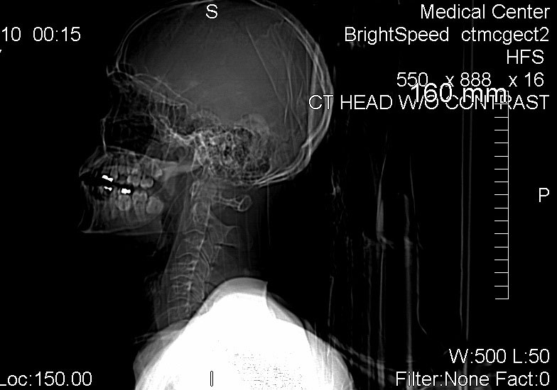
- CT Scan
Nasal Fractures – Broken Nose
The American Academy of Otolaryngology has a comical start to the avoidance of nasal fractures. Because they are commonly caused by two major events, the Academy tells you to:
- Wear protective gear to shield your face when playing contact sports like football
- Don’t get in fist fights
A broken nose is common in society and usually easily treated. If treating this injury, you should look for obstructions to the airway and deformities, as these are the main types of damage.
Occipital Skull Fractures
The occipital bone is the plate shaped base of your skull. The occipital condyle is just above the top of your spine. There are two types of occipital fractures, which include Type I and Type II. They are usually treated conservatively. Expect neck stabilization using HALO traction or Philadelphia collar.
Occipital skull fractures are very serious bone breaks. They can cause death and are often associated with C1 spine injuries. Accordingly, paralysis is a concern. Every occipital skull fracture should seek immediate medical treatment.
Orbital Skull Fractures – Broken Eye Bone
Three percent of all emergency department visits have to do with eye injuries. Broken orbital bones are typically a result of auto accidents where an occupant is not wearing a seat belt. They are the most common type of maxillofacial trauma.
One issue with orbital skull fractures is children. These broken eye bones are often overlooked with kids injured through various items be they accidents or play.
There are three categories of severity for orbital fractures. Treatment for these range from nose surgery, whatsoever, to a major trauma incident. The types are:
- Trapdoor fractures – Caused by low force
- Medial blow-out fractures – Caused by intermediate force
- Lateral blow-out fractures – Caused by high force
The primary goal of an orbital fracture treatment is the prevention of the loss of an eye or to minimize the problems with eye sight. The secondary goal to orbital fracture is disfigurement or a wrong positioning of the eyeball and surrounding tissue.
Zygomatic (Cheekbone) Breaks
A broken cheekbone can be treated easily. Between ten and fifty percent of all these bone breaks require no surgery. Rather, the hospital or physician observes regularly (usually weekly) as the broken cheekbone heals.
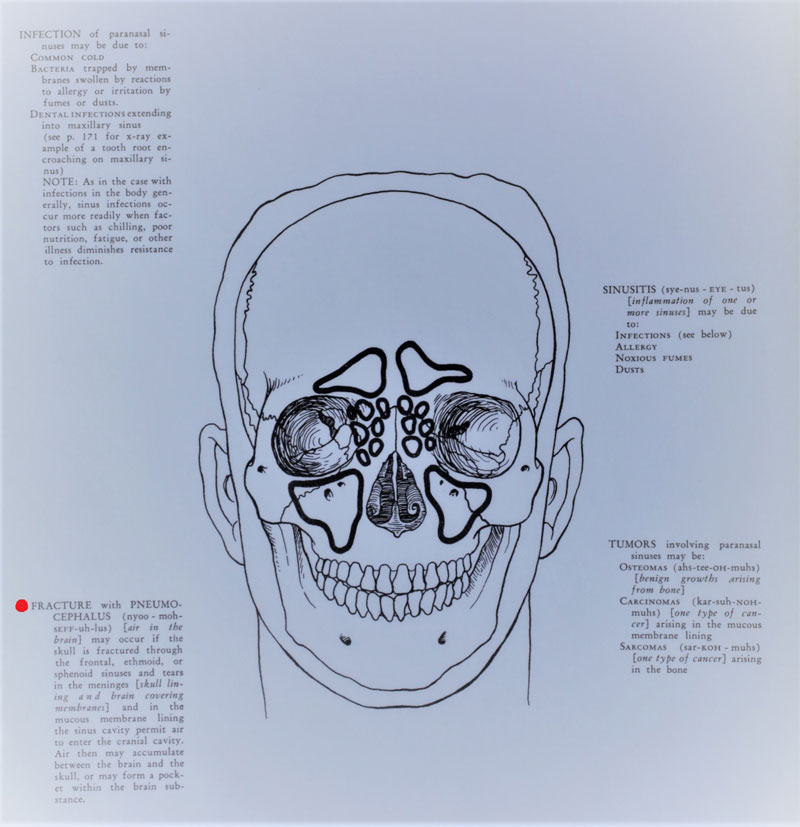
Bleeding through the nose is a particular problem with these zygomatic breaks. There are times when the orbital walls around the eye can be harmed. Air can be forced into the retrobulbar space and cause pain and even the loss of eye sight.
How common are skull fractures?
1.7 million people in the United States sustain a head injury each year. Approximately 37 percent of these injuries include a skull fracture. More conservative studies show that skull breaks are as rare as 42,409 people in the United States per year.
The most common broken bones in the skull are:
- Parietal Bone
- Temporal
- Occipital
- Frontal Bone Break
Broken skull bones can be broken in several ways. The most common types of skull fracture breaks are:
- Linear fractures
- Depressed fractures
- Basilar fractures
Men are more likely to have a fractured skull then women by far. The statistics show:
- 79% Men
- 21% Female
Skull fractures are also a product of youth. Age pans out as follows:
- 29 years old equals average age.
- 75 years old and older equals 4% of skull fractures
Trauma to the head is the most common cause of a broken skull bone. The most common causes of skull fractures are:
- Gun shots
- Auto accidents
- Motorcycle accidents
- Bicycle accidents
- Construction injuries
- Slipping and falling
- Pedestrian accidents
- Assault
Treatment usually includes hospitalization. This produces the following statistics:
- 78% equals hospitalization
- Average length equals 85% one day, 95% two days
Skull Fracture Symptoms
Skull fracture symptoms are observations identified by treating physicians in a number of ways. Clinical examinations and objective tests are the two categories. These pan out as follows:
Clinical Examination:
- Bruising in the face
- Bleeding through the ears and nose
- Fluid leaking from the ears (CSF otorrhea)
- Pain at the impact sight
- Redness at the impact sight
- Warm at the impact sight
- Headaches
- Nausea
- Vomiting
- Dizziness
- Blurred vision
- Restlessness
- Irritability
- Loss of balance
- Neck stiffness
- Eyes not reacting to the light
- Confusion
- Excessive drowsiness
- Fainting or loss of consciousness
Objective Clinical Tests:
- CT Scan – CAT Scan (most common)
- X-Rays
- MRIs
- Long-Term Complications from Skull Fractures
Broken head bones and other skull fractures cause a number of long-term issues. Medical treatment seeks to alleviate these. Lawsuits and other insurance claims from skull fractures include these as items of damages:
- Infection
- Brain damage (TBI)
- Cognitive impairment
- Behavioral changes
- Contusions (bruising)
- Bleeding into one’s brain (cerebral hematoma)
- Brain swelling
- Paralysis
- Coma
- Death
- Diffuse Axonal injuries
- Brain bruising
- Brain lesions
- Meningitis
- Parietal lobe damage
- Scalp damage
- Brain hemorrhage
- Seizures
Treatment of Skull Fractures
Skull fractures can be treated based on the symptoms they cause. Often, they require no formal treatment and the victim can be discharged to go home. Other skull fractures require hospital admission for observation. This is especially true if the victim is an infant. Antibiotics are typically not prescribed unless it is an open fracture, which may also require a tetanus shot. One in every five skull fractures require seizure medication. Surgery is the most serious of the treatments and is typically required if cerebral spinal fluid is leaking from the ears or nose or into the brain causing a pressure build up.
 How to choose the best skull fracture lawyer?
The best Kansas City bone attorneys will know the distinctions, treatment, and damage elements for a skull fracture accident. This is more important than a typical personal injury case or auto accident injury attorney. This is because skull fractures present unique items of damages. These damages will need to be sufficiently proven to get full value for a case.
Our advice is to read and familiarize yourself with particulars of your injuries, its treatment, and long-term complications. Then discuss with the attorneys you are considering how to get these into evidence and best prove it for full case value. The best broken bone attorneys will know experts and the best way to present this evidence. The average or the less than Skull Fracture Lawyer will give an excuse or some other broad statement but not actually answer the questions. This is an area where targeted experience and specific success pays a diffidence in money compensation to the victim.

